


Angiography
Angiography is a medical procedure used to visualize and examine blood vessels in various parts of the body, particularly in the heart and brain. It is a type of imaging test that helps doctors see how blood is flowing through the blood vessels and if there are any blockages or abnormalities.
During angiography, a contrast dye is injected into the blood vessels, which makes them visible on X-ray images or other imaging techniques. The contrast dye helps highlight the blood vessels, making it easier for doctors to identify any issues.
There are different types of angiography depending on the area of the body being examined:
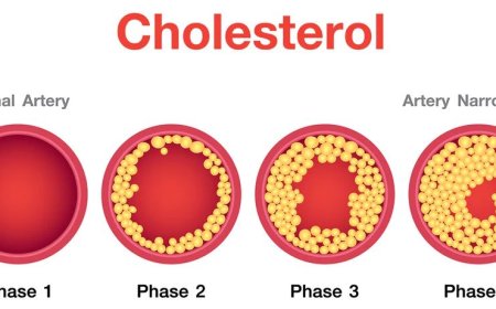
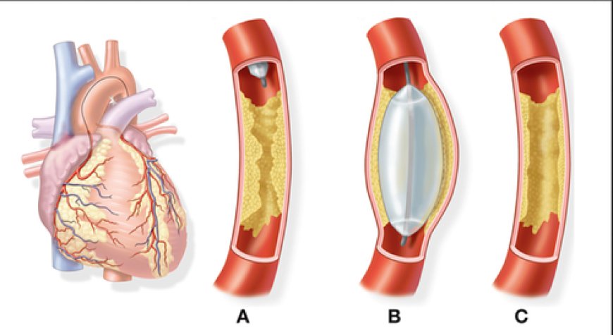
Coronary Angiography: This is a common type of angiography used to examine the blood vessels of the heart. It helps detect blockages or narrowing in the coronary arteries, which supply oxygen-rich blood to the heart muscle.
Cerebral Angiography: Also known as cerebral arteriography or brain angiography, this type of angiography examines the blood vessels in the brain. It is used to identify conditions like aneurysms, arteriovenous malformations (AVMs), and other abnormalities.
Peripheral Angiography: This type of angiography is used to examine blood vessels in the arms, legs, and other parts of the body. It helps diagnose peripheral arterial disease (PAD) and assess blood flow in these areas.
Angiography is performed by a specialized medical professional called an interventional radiologist or an interventional cardiologist. It is typically done in a hospital's special imaging suite or catheterization lab.
Explanation of the procedure and steps in angiography
Step 1: Preparation
- Before the angiography, you will be asked to change into a hospital gown, and a small needle will be inserted into your arm or hand to deliver medications and fluids during the procedure.
Step 2: Numbing the Area
- You will lie on a special table, and the doctor will clean and numb the area where they will insert a tiny tube called a catheter. The catheter is very thin, like a spaghetti noodle!
Step 3: Inserting the Catheter
- The doctor will gently insert the catheter through your skin into a blood vessel, usually in your leg or arm. You may feel a bit of pressure, but it shouldn't hurt.
Step 4: Guiding the Catheter
- Using X-ray guidance, the doctor will gently thread the catheter through the blood vessels until it reaches the area of interest, like the heart or brain.
Step 5: Contrast Dye
- Once the catheter is in place, the doctor will inject a special dye through the catheter. This dye helps highlight the blood vessels on X-ray images, making them visible.
Step 6: X-ray Pictures
- As the dye moves through the blood vessels, the doctor will take X-ray pictures in real-time. These images show how blood flows through your blood vessels and help identify any blockages or narrowings.
Step 7: Catheter Removal
- After the procedure is complete, the doctor will gently remove the catheter, and pressure will be applied to the insertion site to prevent bleeding.
Step 8: Recovery
- You will be taken to a recovery area to rest for a short while. The healthcare team will monitor you to ensure you are doing well after the procedure.
Step 9: Going Home
- Once you're feeling better and the doctor gives the green light, you can usually go home on the same day.
Diagnosing and Treating Vascular Conditions:
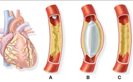
Angiography plays a critical role in diagnosing various vascular conditions, guiding treatment decisions, and assessing the effectiveness of interventions. For instance, in coronary angiography, doctors can identify blockages in heart arteries and perform angioplasty to open them, restoring blood flow to the heart muscle.
Risks and Benefits of Angiography
Benefits:
Accurate Diagnosis: Angiography allows doctors to see inside your blood vessels in real-time, helping them pinpoint the exact location of blockages, narrowings, or other problems. This precise diagnosis is crucial for effective treatment planning.
Guiding Treatment Decisions: By providing detailed images of your blood vessels, angiography helps doctors determine the best treatment options. Whether it's opening blocked arteries with angioplasty or planning surgery for an aneurysm, angiography ensures tailored and targeted interventions.
Real-time Visualization: During the procedure, doctors can immediately see how blood flows through your vessels, which is particularly helpful during emergency situations like a heart attack or stroke.
Minimally Invasive: Angiography is usually less invasive than traditional surgery. The use of a thin catheter means smaller incisions and quicker recovery times.
Risks:
Allergic Reactions: Some people may be allergic to the contrast dye used during angiography. However, these reactions are rare, and medical teams are well-prepared to handle any emergencies.
Bleeding and Bruising: After the procedure, you may experience mild bleeding or bruising at the site where the catheter was inserted. These usually heal on their own.
Blood Vessel Injury: There is a small risk of injury to the blood vessels during the insertion of the catheter. However, highly trained medical professionals perform the procedure to minimize such risks.
Rare Complications: Although extremely uncommon, there is a slight chance of more serious complications, such as blood clots or infections. However, these are exceptionally rare.
What's Your Reaction?
 Like
0
Like
0
 Dislike
0
Dislike
0
 Love
0
Love
0
 Funny
0
Funny
0
 Angry
0
Angry
0
 Sad
0
Sad
0
 Wow
0
Wow
0